emergency medicine
A 57-year-old woman with metastatic breast cancer
It's Metastatic Breast Cancer Quiz Time with Med-Challenger!
Breast fat necrosis can mimic cancer, including skin retraction, but is benign and not a cancer risk. High-yield board review with distractors.
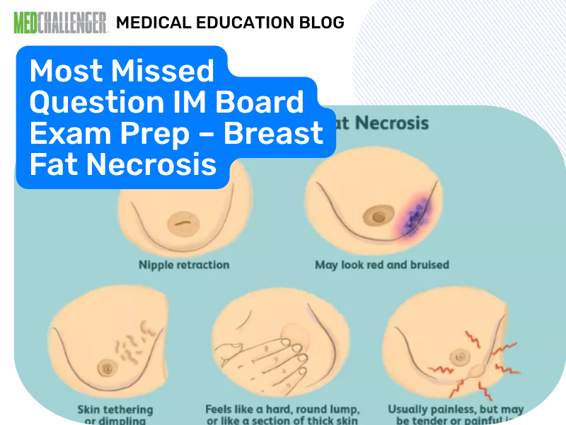
Fat necrosis is a benign breast condition that can mimic cancer clinically and on imaging, including causing skin retraction.

During a routine annual physical exam a 43-year-old woman is found to have a mass in the right breast. Mammography reveals a suspicious mass. A biopsy of the lesion reports fat necrosis.
Which of the following statements concerning fat necrosis of the breast are true?
Answer Options:
Fat necrosis commonly follows trauma, surgery, radiation, anticoagulation-related hematoma, or inflammation—and it can feel and look like breast cancer. On boards, the key is recognizing that skin tethering/retraction does not automatically equal malignancy; benign entities like fat necrosis can do this.
Another major exam point: fat necrosis is benign and is not a premalignant lesion. If imaging is suspicious or there’s clinical–radiologic discordance, the “standard pathway” reflected in ACR BI-RADS/NCCN diagnostic algorithms is to pursue appropriate tissue diagnosis—consistent with this stem already having biopsy confirmation.
| Option | What It Tests / Implies | Why It’s Wrong Here |
|---|---|---|
| Fat necrosis results in a diffuse, poorly defined mass y | Malignancy-like “ill-defined/diffuse” infiltrative mass | Fat necrosis typically presents as a localized palpable lesion; while imaging can look spiculated, the classic teaching is not “diffuse poorly defined mass” as a defining feature. |
| Fat necrosis may be accompanied by skin retraction | Clinical mimicry of carcinoma | Correct: fat necrosis can cause skin retraction/tethering and mimic cancer clinically and radiographically. |
| Fat necrosis confers a twofold increased risk of subsequent infiltrating lobular carcinoma | Whether fat necrosis is a premalignant/risk lesion | Incorrect: fat necrosis is benign and does not confer a known increased risk of carcinoma. |
| Most women relate the tender breast mass to a breast trauma in the previous two to four weeks | Recall of antecedent trauma | Incorrect: many patients do not recall trauma (or the timing is variable); history of trauma is supportive but often absent. |
A palpable breast mass with skin retraction can still be benign (e.g., fat necrosis), but suspicious imaging/discordance must follow BI-RADS/NCCN diagnostic pathways to tissue diagnosis.
The item exploits a common heuristic—“retraction means cancer”—and tests whether you can keep a broad differential and remember that certain benign processes (notably fat necrosis) can produce carcinoma-like physical and mammographic findings.
A 52-year-old woman develops a firm palpable breast mass 2 months after lumpectomy. Mammogram shows calcifications; ultrasound suggests an oil cyst. Which statement is most accurate?
A 45-year-old has a palpable breast mass with mild skin dimpling. She has a history of blunt breast trauma months ago. Which diagnosis can explain dimpling while still being benign?
A patient asks if biopsy-proven fat necrosis increases her future breast cancer risk. Best response?
A 48-year-old has a new breast lump. Mammogram is BI-RADS 5. She reports recent trauma and you suspect fat necrosis. Best next step?
Which history feature is most consistent with fat necrosis (though not always present)?
How would your diagnostic approach differ between (1) a patient with a new palpable mass and BI-RADS 5 imaging who reports trauma and (2) a patient with biopsy-proven fat necrosis and reassuring concordant imaging?
Q1: Can fat necrosis cause skin dimpling or retraction similar to cancer?
A: Yes. Board exams (e.g., ABIM-style questions) expect recognition that fat necrosis can create fibrosis and tethering, mimicking carcinoma on exam.
Q2: Does fat necrosis increase the risk of future breast cancer?
A: No. It’s a benign, nonproliferative process; malignancy risk counseling should reflect that it does not itself confer elevated risk.
Q3: If trauma history suggests fat necrosis, can you skip biopsy when imaging is suspicious?
A: No. The exam-relevant rule is that suspicious imaging (e.g., BI-RADS 4/5) follows diagnostic algorithms (ACR BI-RADS/NCCN) that typically require tissue diagnosis.
Q4: Why is this a “most missed” concept?
A: Many test-takers equate retraction with malignancy and overvalue trauma recall; exams test whether you know benign mimics still require proper diagnostic workup.
This question appears in Med-Challenger Internal Medicine Review with CME
Try for free and save. Ace your exams and meet your CME/MOC requirements for just $35 a month!



No matter your program, no matter the size, Med-Challenger for Groups and Institutions can better prepare your program or group, fulfill industry requirements, and increase test scores.
For personal medical education that includes board's prep, MOC, and CME requirements, Med-Challenger has you covered in Family Medicine, Emergency Medicine, Internal Medicine, Pediatrics, Pediatric Emergency Medicine, OBGYN, Physician Assistants, and Nurse Practitioners.
It's Metastatic Breast Cancer Quiz Time with Med-Challenger!
It's Family History of Breast Cancer Quiz Time with Med-Challenger!
Staffing Crisis Costing Millions AI Powered Supplemental Course Info AI Detects Cancer Better Refer a Friend and Save Healthcare is a Bludgeon GLP-1...