Quick take: a pediatric EMT/PEM board-style question on post‑intubation chest x‑ray interpretation and ET‑tube position in a 13‑year‑old with myotonic dystrophy. Read the question, answer rationale, and remediation links below.
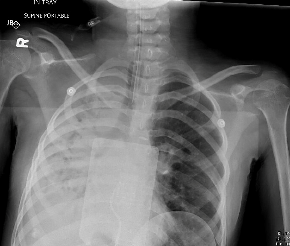
A 13 yo adolescent girl with history of myotonic dystrophy presents with a 2 day history of cough, nasal congestion, and several episodes of non-bloody nonbilious emesis presents in respiratory distress. On examination her oxygen saturation is 80%, she is sleepy and minimally responsive to painful stimuli. She is intubated with fentanyl, midazolam, and rocuronium. The post-intubation chest x-ray is shown below. What is the most likely cause of the chest x-ray findings.
 Image courtesy of Ashley Keilman, MD
Answer Options:
- Aspiration pneumonia
- Pleural effusion
- Pneumothorax
- Right mainstem intubation
The correct answer is 1, aspiration pneumonia. The question is missed surprisingly often, with the most often incorrect answer being pleural effusion. There’s a classic location, and air-filled bronchi in the opacity. Diaphragm silhouettes are intact. Not fluid in the pleural space. No real ‘test trick’ is used here. 🙂
Why This Question Is Often Missed
- Supine portable film pitfalls: Learners often misinterpret dependent consolidation on a supine film as a pleural effusion, since fluid-layering signs (e.g. meniscus) are not obvious.
- Anchoring on tube position: The presence of an ETT and sedatives tempts some to jump to “right mainstem intubation” without carefully assessing radiographic lung findings.
What the Distractors Indicate
| Option |
What It Tests / Implies |
Why It’s Wrong Here |
| Aspiration pneumonia |
Recognition of dependent consolidation in intubated patient |
Correct: Patchy, gravity‑dependent opacity in superior right lower lobe on a supine film is classic for aspiration. |
| Pleural effusion |
Ability to identify pleural fluid (meniscus, blunting) |
No meniscus or costophrenic blunting; opacity is lobar, not pleural‑based. |
| Pneumothorax |
Identification of extra‑pulmonary air (absent markings) |
Lung markings extend to periphery; there is no lucent line or absence of vascular markings. |
| Right mainstem intubation |
Correlation of ETT depth with unilateral hyperinflation |
Both lungs contain air; the tube tip lies mid‑tracheal above the carina. |
High-Yield Pearl
On a supine portable CXR, aspiration pneumonia often presents as gravity‑dependent consolidation in the superior segment of the lower lobes, which can mimic effusion—look for preserved lung volume and bronchograms.
Core Learning Objectives
- Radiographic recognition of aspiration pneumonia in intubated pediatric patients on supine portable films.
- Differentiation of common post‑intubation complications (e.g., mainstem intubation, pneumothorax, effusion) on chest radiographs.
The “Test Trick” at Play
Examiners exploit the supine portable film’s tendency to obscure classic signs (e.g., meniscus for effusion, clear fluid levels) and couple it with the distraction of the endotracheal tube to lure candidates toward tube‑related or pleural‑based diagnoses rather than true lobar consolidation.
Additional Peds EM Practice Questions and Remediation
Pediatric Emergency Medicine Practice Question 1
A 10‑year‑old boy with cerebral palsy is intubated emergently after aspiration of gastric contents. Supine CXR shows patchy opacity in the left lower lung zone with air bronchograms. What is the most likely cause?
A. Aspiration pneumonia
B. Pulmonary contusion
C. Atelectasis from mucus plugging
D. Left pleural effusion
Pediatric Emergency Medicine Practice Question 2
A 15‑year‑old with status epilepticus is sedated, intubated, and develops fever. Supine CXR 48 h later shows dense consolidation in the right upper lobe. Which factor most predisposed him?
A. Tube‑feed aspiration
B. Supine positioning
C. Prolonged seizure activity
D. High-dose benzodiazepines
Pediatric Emergency Medicine Practice Question 3
After intubation in the ED, a 12‑year‑old’s CXR shows unilateral hyperlucency on the right and mediastinal shift to the left. The ETT tip is 2 cm above the carina. What complication does this represent?
A. Subcutaneous emphysema
B. Tension pneumothorax
C. Right mainstem intubation
D. Pulmonary embolism
Pediatric Emergency Medicine Practice Question 4
A 9‑year‑old post‑intubation CXR shows opacity in the left lung base with loss of diaphragmatic contour but normal lung volume. Which finding confirms pneumonia over effusion?
A. “Spine sign” on lateral view
B. Silhouette sign of the diaphragm
C. Air bronchograms
D. Lateral decubitus free fluid
Pediatric Emergency Medicine Practice Question 5
A child with a seizure disorder is found unresponsive in vomitus, intubated, and CXR reveals dependent consolidation in superior segments of both lower lobes. Best next step?
A. Bronchoscopy
B. Diuretic therapy
C. Empiric antibiotics covering anaerobes
D. Immediate chest tube insertion
Mini Case Discussion Prompt
Compare the radiographic features and management strategies of aspiration pneumonia versus right mainstem intubation in an intubated pediatric patient on a supine portable film—what clinical clues and positioning changes could help differentiate them?
This question appears in Med-Challenger Pediatric Emergency Medicine 3rd Edition Exam Review with CME
Try for free and save. Ace your exams and meet your CME/MOC requirements.